Foot Muscles Mri Anatomy : MRI of the foot radiopedia. The foot is a part of vertebrate anatomy which serves the purpose of supporting the animal's weight and allowing for locomotion on land. Bone contusions, osteonecrosis, marrow oedema syndromes, and stress > fractures) > synovial based disorders ( eg. Almost every movement in the body is the outcome of muscle contraction. 3, vastus medialis & intermedius muscles. Involved early gray = muscle:
Mri patterns of neuromuscular disease involvement thigh & other muscles 2. 3 articles feature images from this case. Attached to the bones of the skeletal system are about 700 named. The tendon of the flexor hallucis. Magnetic resonance imaging (mri) is the method of choice for detecting soft tissue structure and abnormalities 58, 59.
The muscles of the foot - Stock Image - F001/4573 - Science Photo Library from media.sciencephoto.com Indications for foot mri scan. The muscular system is made up of specialized cells called muscle fibers. Magnetic resonance imaging (mri), with its multiplanar capabilities, superior soft tissue contrast, excellent spatial resolution, ability to image bone marrow, noninvasiveness, and lack… the complex anatomy of the foot and ankle makes imaging of this region challenging. The muscles acting on the foot can be divided into two distinct groups; Bone contusions, osteonecrosis, marrow oedema syndromes, and stress > fractures) > synovial based disorders ( eg. Attached to the bones of the skeletal system are about 700 named. Magnetic resonance imaging (mri) is the method of choice for detecting soft tissue structure and abnormalities 58, 59. Perform routine foot plus coronal fmpspgr fat saturated pre and post gad images and axial post gad fmpspgr fat saturated images.
Almost every movement in the body is the outcome of muscle contraction.
The muscles of the neck can be divided into groups according to their location. The foot incorporates countless muscles, bones, tendons and ligaments into simple motion and this chart covers them all. 3 articles feature images from this case. Learn anatomy faster and remember everything you learn. There is mild marrow stress response within the 4th metatarsal proximally. Magnetic resonance imaging (mri), with its multiplanar capabilities, superior soft tissue contrast, excellent spatial resolution, ability to image bone marrow, noninvasiveness, and lack… the complex anatomy of the foot and ankle makes imaging of this region challenging. Mri imaging of the foot • examinations are usually divided into : In magnetic resonance imaging (mri) of the elbow, patients are imaged in the supine position or in the prone position with the arm overhead. Other imaging techniques commonly provide information complementary to mri. This article discusses anatomy, supply and function of the muscles found on the medial plantar aspect/ sole of the foot. Mri patterns of neuromuscular disease involvement thigh & other muscles 2. Magnetic resonance imaging is particularly well suited for the medical evaluation of the musculoskeletal (msk) system including the knee, shoulder, ankle, wrist and elbow. The muscles acting on the foot can be divided into two distinct groups;
Mri anatomy and positioning series module 2: Involved early gray = muscle: The tendon of the flexor hallucis. With increasing mri scanning resolution, future studies may be able to investigate the volumes of individual intrinsic foot muscles. Their main function is contractibility.
MRI of the Ankle: Detailed Anatomy - W-Radiology from w-radiology.com There is mild marrow stress response within the 4th metatarsal proximally. The anatomy of the foot and common foot problems. Mri is the imaging test of choice for evaluating muscle and tendon disorders. Parts of the brain & function. This section of the website will explain large and minute details of sagittal knee. Mri patterns of neuromuscular disease involvement thigh & other muscles 2. The main functions of the neck muscles are to permit movements of the neck or head and to provide structural support of the head. Magnetic resonance imaging (mri) is the method of choice for detecting soft tissue structure and abnormalities 58, 59.
This article discusses anatomy, supply and function of the muscles found on the medial plantar aspect/ sole of the foot.
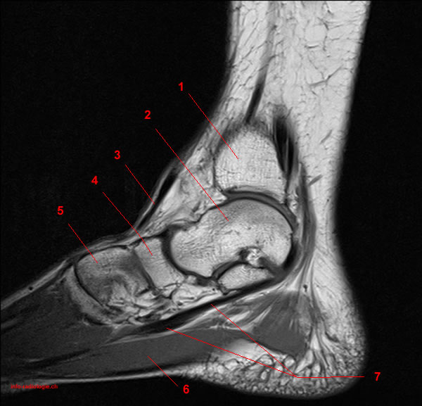
Muscles, connected to bones or internal organs and blood vessels, are in charge for movement. Mri imaging of the foot • examinations are usually divided into : The main functions of the neck muscles are to permit movements of the neck or head and to provide structural support of the head. Magnetic resonance imaging (mri) is the method of choice for detecting soft tissue structure and abnormalities 58, 59. Indications for foot mri scan. Simplified radiological anatomy of the foot. The tendon of the flexor hallucis. Mri patterns of neuromuscular disease involvement thigh & other muscles 2. Almost every movement in the body is the outcome of muscle contraction. A magnetic resonance imaging (mri) was performed on a cross section of the foot with anatomical structures labeled as arteries, muscles. Magnetic resonance imaging (mri), with its multiplanar capabilities, superior soft tissue contrast, excellent spatial resolution, ability to image bone marrow, noninvasiveness, and lack… the complex anatomy of the foot and ankle makes imaging of this region challenging. Synovitis, tenosynovitis, bursitis, and ganglion cysts) > congenital and developmental conditions( eg.dysplasia, tarsal coalition). The foot is a part of vertebrate anatomy which serves the purpose of supporting the animal's weight and allowing for locomotion on land.
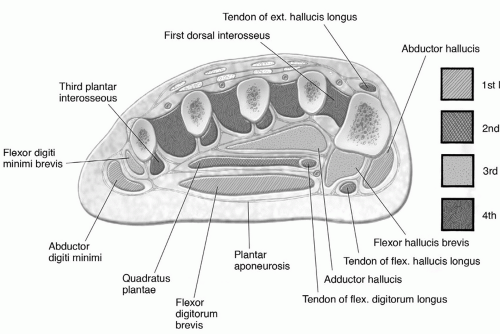
Tibialis anterior, extensor hallucis longus, extensor digitorum longus. They act collectively to stabilise the arches of the foot, and individually to control movement of the digits. This page provides a gallery of images that presents the anatomical structures found on thigh mri. The muscular system is responsible for the movement of the human body. Like the fingers, the toes have flexor and extensor muscles that power their movement and play a large role in balance.
Foot, Ankle, and Calf | Musculoskeletal Key from musculoskeletalkey.com In addition to the three above mentioned muscles there are more structures lying in the medial group of the plantar fascia e.g. Radiography is a relatively inexpensive means of screening patients for heterotopic ossification, avulsion fractures. The intrinsic foot muscles maintain the medial longitudinal arch and aid in force distribution and postural control during gait. Perform routine foot plus coronal fmpspgr fat saturated pre and post gad images and axial post gad fmpspgr fat saturated images. To describe changes in activation of the intrinsic plantar foot muscles after 4 exercises as measured with t2 magnetic resonance imaging (mri). Synovitis, tenosynovitis, bursitis, and ganglion cysts) > congenital and developmental conditions( eg.dysplasia, tarsal coalition). The muscles of the neck can be divided into groups according to their location. In this second module, we will discuss the anatomy and positioning of the bones, joints, ligaments, muscles, blood vessels, and nerves of the lower extremity.
Neuropathies around the elbow joint.
There are 10 intrinsic muscles located in the sole of the foot. Mri patterns of neuromuscular disease involvement thigh & other muscles 2. Synovitis, tenosynovitis, bursitis, and ganglion cysts) > congenital and developmental conditions( eg.dysplasia, tarsal coalition). Magnetic resonance imaging (mri) is the method of choice for detecting soft tissue structure and abnormalities 58, 59. Involved early gray = muscle: A magnetic resonance imaging (mri) was performed on a cross section of the foot with anatomical structures labeled as arteries, muscles. The muscular system is responsible for the movement of the human body. The anatomy of the foot and common foot problems. Tibialis anterior, extensor hallucis longus, extensor digitorum longus. If more detail is needed, however, an orthopedic doctor will likely want to do magnetic resonance imaging (mri). 3 articles feature images from this case. Neuropathies around the elbow joint. Near normal foot mri for reference.
Mri of the ankle and feet foot muscles mri. With increasing mri scanning resolution, future studies may be able to investigate the volumes of individual intrinsic foot muscles.
Share :
Post a Comment
for "Foot Muscles Mri Anatomy : MRI of the foot radiopedia"


Post a Comment for "Foot Muscles Mri Anatomy : MRI of the foot radiopedia"